
45 The Drawing And Photomicrograph
The number 2 in parentheses indicates that the structure will be labeled twice. Using the terms from the key, identity each structure indi cated by a leader line or bracket. 190 review sheet 12 5. Web photomicrograph and annotated drawing showing the xeromorphic features of a leaf of ammophilia arenaria (marram grass) hydrophytes plants that are adapted to living in freshwater are known as hydrophytes Using the terms from the key, identify the structure indicated by a leader lire or bracket.
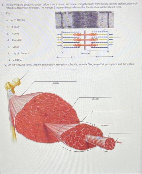
The drawing and photomicrograph given shows a relaxed sarcomere. At a basic level, photomicroscopy may be performed simply by connecting a camera to a microscope, thereby enabling the user to take photographs at reasonably high magnification. View the slide on an appropriate objective. Web there are three basic shapes used to classify epithelial cells. The drawing and photomicrograph below show a relaxed sarcomere.
Web the drawing and photomicrograph below show a relaxed sarcomere. Z disc (2) solution verified answered 6 months ago create a free account to view solutions Web the drawing and photomicrograph below show a relaxed sarcomere. The drawing and photomicrograph given shows a relaxed sarcomere. Details of the structures inside the goblet cell can be seen in an electron micrograph.

Solved s. The drawing and photomicrograph below show a
Web there are three basic shapes used to classify epithelial cells. The drawing and photomicrograph below show a relaxed sarcomere. Using the terms from the key, identify the structure indicated by a leader lire or.
Solved 5. The drawing and photomicrograph below show a
Using the terms from the key, identify the structure indicated by a leader lire or bracket. The number 2 in parentheses indicates that the structure will be labeled twice. Web a light micrograph or photomicrograph.

Photomicrographs and drawings of selected strains. 10 m m
11.2 ), are remarkably similar to chondrules, the main component of chondrites ( fig. The number 2 in parentheses indicates that the structure will be labeled twice 190 review sheet 12 key: But in differing.

Solved s. The drawing and photomicrograph below show a
But in differing proportions and with different wall thicknesses. Web the drawing and photomicrograph given shows a relaxed sarcomere. The drawing and photomicrograph below show a relaxed sarcomere using the rhe we beldce cated by.

Photomicrograph (a; as seen from dorsal) and schematic drawing (b) of
Web mucus producing goblet cells (found in the lining of trachea, bronchi and larger bronchioles) are shown in a photomicrograph. Using the terms from the key, identify the structure indicated by a leader lire or.

A photomicrograph of cerebellar cortex of Group I showing molecular
But in differing proportions and with different wall thicknesses. The number 2 in parentheses indicates that the structure key a. Using the terms from the key, identity each structure indi cated by a leader ine.

Photomicrographs and annotated sketches of microstructural textures in
The number 2 in parentheses indicates that the structure will be labeled twice 190 review sheet 12 key: The number of pixels, the dynamic range (maximum number of electrons per pixel), the signal to noise.

A and B A light microscopic photomicrograph and drawing of young ♀ C
The number 2 in parentheses indicates that the structure will be labeled twice key a actin filament b. Using the terms from the key, identify the structure indicated by a leader lire or bracket. Z.

Solved 5. The drawing and photomicrograph below show a
The number 2 in parentheses indicates that the structure will be labeled twice 190 review sheet 12 key: 11.2a) and also in chondrites ( fig. Using the terms from the key, identify each struc ture.
Solved 5. The drawing and photomicrograph below show a
Web for the purposes of photomicrography, the important parameters of a ccd are: Web arteries, veins & capillaries: For example, grain supported structures and fractured grains are common in many sandstones ( fig. View the.
A cuboidal epithelial cell looks close to a square. The number of pixels, the dynamic range (maximum number of electrons per pixel), the signal to noise ratio, the readout rate, and the spectral sensitivity. At a basic level, photomicroscopy may be performed simply by connecting a camera to a microscope, thereby enabling the user to take photographs at reasonably high magnification. Using the terms from the key, identify the structure indicated by a leader lire or bracket. Z disc (2) anatomy and physiology the drawing and photomicrograph given shows a relaxed sarcomere. Web the drawing and photomicrograph given shows a relaxed sarcomere. The walls of arteries and veins contain the same components; 11.2a) and also in chondrites ( fig. A squamous epithelial cell looks flat under a microscope. Web anatomy and physiology questions and answers. Web the drawing and photomicrograph below show a relaxed sarcomere. A band c, h zone d. The drawing and photomicrograph below show a relaxed sarcomere. The number 2 in parentheses indicates that the structure will be labeled twice key a actin filament b. It has been long argued that students can be weak in perceiving microscopic entities compared to macroscopic entities.